Saturday 3 November 2018

Pneumonia


It is lower respiratory tract infection that cause inflammation of alveoli sacs.

- Germs like bacteria , virus and fungi attack the system. Alveoli , Bronchioles helps to gas exchange. Normal respiratory system fight of the germs by taking air through nose but certain conditions. When Alveoli sac get inflamed  and it starts fill with full of fluid , there is aggregation of WBCs and RBCs and bacteria which impair gas exchange.

Risk factors- 


1. Influenza -   influenzai highly contagious viral infection which spread easily from person to person. Influenza is most common cause of Pneumonia commonly in young child , pregnant mother.

2. Weak immune system- this also leads to pneumonia due to low immunity bacteria get easily multiply.

3. HIV infection - This leads to opportunistic infection for pneumonia. In HIV infection there is low immunity which leads to pneumonia.

4. Medications which spress autoimmune system.

5. Decreased consciousness and Increased risk of aspirations.

6. Chronic obstructive pulmonary disease.(COPD).

7. Post- op abdominal and chest surgery. After surgery there is chances of getting bacterial infection which may cause pneumonia.

8. Respiratory acidosis - in this lungs retain carbon dioxide and can't get oxygen to blood which cause hypoxemia. And decrease in PH.

Causes-


Most common causes pneumonia are 

1. streptococcal pneumoniae.
2. Mycoplasma pneumonia.

Virus - 
1. Influenza

Types-


1. Community Acquired- 

This type of infection occur patient get germs outside the health care settings.

2. Hospital acquired-

This type of infection occurs when patient in hospital settings and mainly occur in mechanical ventilation. This infection occurs 48-72 hrs after admission.

Diagnose- 

1. Notice on auscultation a coarse crackles bronchi or bronchioles breath sound due to lung consolidation.

2. X-ray

3. Sputum culture.

Sign and symptoms-


1. Productive cough.
2. Pain in chest.
3. Unusual breath sound.
4. Altered lab values e.g. Increased partial pressure of carbon dioxide and incrIncre WBCs.
5. Unusual breath sound.
6. Mild to high fever.
7. Nausea vomiting.
8. Increased Heart rate and pulse rate.
9. Activity intolerance.

Treatment-


Medications- 


1. Vancomycin- used in severe cases and can treat resistance bacteria. Most common side effect may have oto- toxicity.

2. Macrolide- Narrow spectrum and kill gram +ve bacteria.

3. Tetracycline- Broad spectrum not used in pregnancy.

4. Fluroquinolone - Broad spectrum and have side effect of frequent diarrhea , tendon rupture.

5. Cephalosporin.

6. Pencillin.

Supportive care-


1. Oxygen therapy.
2. Oral rehydration therapy.
3. Iv fluids.

Patient care-

1. Monitor lung sound impairment.
2. Monitor vital sign. Especially saturation and pulse rate.
3. Assess color of skin for cyanosis.
4. Monitor ABG results.
5. Collect sputum for test as ordered.
6. Provide suctioning if needed.
7. provide breathing and respiratory exercises.

Education- 


1. Encourage for incentive spirometry.

2. Encourage to hydrated and take 2-3 L/day water to keep secretions thin. And patient with heart failure and renal failure don't give more fluids.

3. Make sure patient is up to date with pneumothorax vaccine every 5 years.

4. Encourage to stop smoking and maintain hygiene.

5. Administer medications as per ordered.


Thursday 1 November 2018

COPD symptoms and treatment


It is a pulmonary disease which cause chronic destruction of airflow from lungs.
There is limited airflow due to the inflamed bronchioles and get narrow with excessive mucous. It also lead to inability to fully exhale.
This disease is irreversible and cases vary from mild to severe . This disease is managed with life style changes and medications.

Causes-


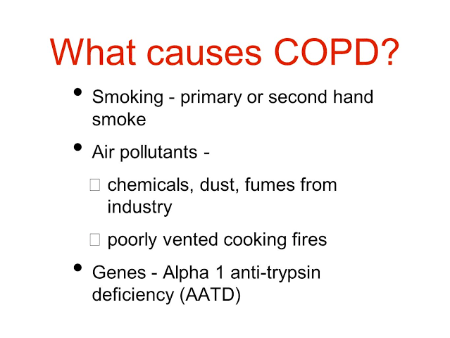
Most common cause is environmental from 
harmful irritants that are Inhaled into lungs e.g smoking.

- this disease happen gradually and most patient strat to notice sign and symptoms in middle age.

Types-


This disease is used now catch all terms for disease that limit air flow.

1. Chronic bronchitis-


This is also called bloating due to cyanosis and edema due to right heart failure. This lead to respiratory acidosis.

-   Cyanosis due to not proper gas exchange. This status starts to compensate by the body by producing more RBCs in the body which leads to  more problems that the blood get thick and pressure on pulmonary artery get Increased and this lead to backflow of the blood from the artery to left atrium which lead to left side heart failure and lead to the edema of extremities.

2. Emphysema (pink puffer)-


It is called pink puffer because body don't have cyanosis. puffer is called because there is hyperventilation for compensation when decreased oxygen and breath rapidly.

In this alevoali loss the elasticity and which leads to not entering of  oxygen and exhale carbon dioxide which leads to lungs hyperinflation and which cause the depression of diaphragm from dome shape to flat. And this lead to compensation by the use of accessory muscles to breath and lead to barrel chest due to which anteroposterior diameter get Increased.

- hyperventilation occur and which does not lead cyanosis and have pink complexion.

Sign and symptoms-


1. Lack of energy due to improper blood supply.
2. Unable to tolerate activities due to lack of energy.
3. Loss of weight due to inadequate nutrition.
4. Dry and productive cough.
5. Accessory muscles usage for breathing most commonly in  emphysem , abnormal lung sound , coarse crackles or wheezing.
6.  Barrel chest due to increase in anterior diameter.
7. Extreme dyspnea.
8. Patient used one fixed position to breath. E.g. use of stick to breath..

Complications-


1. Heart failure.
2. Pneumothorax.
3. Lung infection.
4. Increased risk for lung cancer.

Diagnose- 


1. Spirometry-   to check to which extent  patient can breath and how much volume the lung can hold during inhalation and how much and how fast volume get exhaled.

2. Measuring forced vital capacity.

Patient care-

1. Monitor respiratory system.

2. Monitor lung sound if secretion present than suctioning may required.

3. There may be sputum production( Collect production if ordered for testing). There may be risk for pneumonia.

4. Keep saturation 89-93 % because patient with COPD are stimulated to breath due to low oxygen level rather than high carbon dioxide level. If we give them too much oxygen they start hypoventilating and which leads to carbon dioxide toxicity.

5. Monitor for efforts of breathing and encourage to pursed lip breathing.

6. Administer breathing treatment eg. Nebulization and corticosteroid as prescribed.

Education- 


1. Nutrition need - eat high calories and protiens meals. Maintain fluid intake 2-3 L/ day.

2. Instruct to avoid sick people , irritants hot humid or extreme cold also can affect them.

3. Instruct to stop smoking.

4. Vaccinations should be up to date.

Medications-


1. Corticosteroid- decrease inflammation and decrease mucous production. These are given oral , IV , Inhaled, they are used many times.
These drugs included - 

A. Prednisone.
B. Solumedrol.

2. Bronchodilator-  should used first than corticosteroid if both prescribed to the patient.

- After the use of corticosteroid inhaler patient should rinse mouth because it may cause Thrush.

3. Phosphodiesterase -4 Inhibitors ( Roflumilas)- used for patient with chronic bronchitis and help to decrease COPD exacerbation not bronchodilators. These drugs have side effect that it cause suicide ideation and weight loss.

4. Theophylline - Given orally many times. This is type of bronchodilators and used long term with sever COPD.

5. Short acting bronchodilators- Relax smooth muscles bronchial tubes and are used in emergency conditions for quick relief.

A. Albuterol - Beta 2 blockers.
B. Atrovent - Anticholinergics.

6. Long Acting Bronchodilators- same action as short acting bronchodilators but used for long time .

A. Salmetrol.
B. Spriva.