Saturday, 20 October 2018

Pancreatitis and treatment

Pancreatitis-


Pancreatitis is the inflammation of pancreas. In pancreas there are number of cells that produces enzymes , hormones and when inflammation occur it leads to digestion of the pancreas by it's own enzymes or cause irreversible structural damage to the organ.


Pancreatitis is of two types-

1. Acute pancreatitis
2. Chronic pancreatitis.

What are the functions of pancreas and how they get affected with pancreatitis-

Pancreas have two types of functions-

1. Exocrine by Acinar cells-

It secret inactive enzymes into pancreatic duct these enzymes are-

  • Amylase - breakdown carbohydrates to glucose
  • Protease -  breakdown protiens.
  • Lipase    - breakdown fats
Pancreas have epithelial cells which release bicarbonate.

2.  Endocrine Islets of Langerhans-

It release insulin , glucagon , somatostatin , pancretic polypeptide into blood stream.

These all enzymes are not activated and they become activated when enter into the duodenum which is part of small intestine. These enzymes reach to duodenum via pancreatic duct.


When pancreatitis occur  which cause inflammation and pancreas get swell up. Enzymes become unable to release due to obstruction in path and these enzymes starts to leak surrounding the pancreatic tissues and also in surrounding organs which leads many complications.

Complications included-


1. Chronic pancreatitis leads to damage to isletI of Langerhans and cause low production of insulin which cause diabetes mellitus.

2. Malabsorption   occur due inadequate secretion of enzymes and their release into small intestine. Which leads to improper digestion of food and cause malabsorption.

3. GI pain due to inflammation of tissues.

4. Internal bleeding due to the inflamation 
of blood vessels.

5. Ascitis occur due to leaking of pancreatic secretions into peritoneum.

6. Fibrosis

7. Cyst formation.

8. Respiratory distress.

What Is difference between acute and chronic pancreatitis? -

Acute pancreatitis-


1. Sudden inflammation due to something that triggers the digestive enzymes to activate inside the pancreas.

2. These activated enzymes inside the pancreas starts to digest cells of pancreas. Which leads to swell up of pancreas and inflamed tissues died cause fibrosis , cyst formation  and abscess formation.

3. Due to location of pancreas it leads to inflammation of other organs. E.g respiratory distress etc.

Chronic pancreatitis-


1. Chronic pancreatitis is repeated episode of acute pancreatitis but most common cause is years of alcohol abuse.

2. It is irreversible change to pancreas.

3. There is complete Loss of endocrine and exocrine cells functions.

4. Complete damage to pancreatic ducts functions.

What are causes of acute and chronic pancreatitis? -


Acute pancreatitis- 

Mainly two causes are dominant-

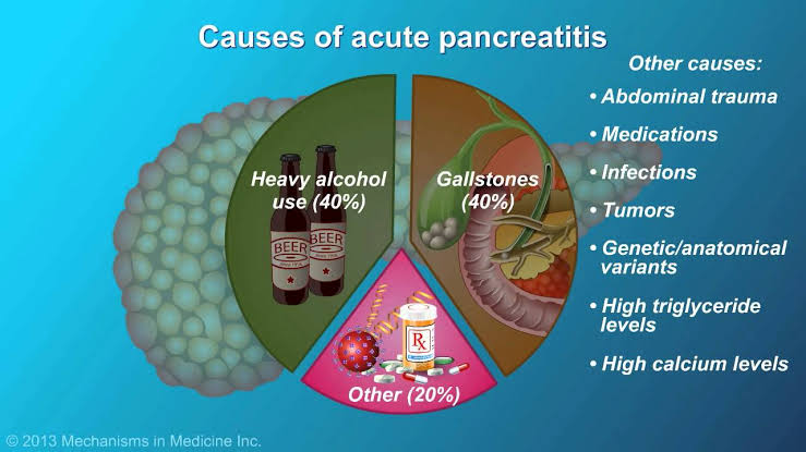
1. Gallstones- hardened deposit of undissolved cholesterol salt or bilirubin that can block common bile duct cause reverse transport of enzymes. These enzymes get activated and start to digest it's own cells in pancreas.

2. Alcohal - leads to damage of the cells of pancreas (Acinar cells) , pancretic ducts and lead to the activation of enzymes.
Other-

  • Abdominal trauma.
  • Medications.
  • Infections.
  • Tumors.
  • Genetics.
  • High triglycerides level.
  • High calcium level.

Chronic pancreatitis -


1. Long term alcohal abuse- recurrent inflammation and damage to the structure of pancreas occur which leads to the chronic pancreatitis.

2. Cystic fibrosis- there is lack of CFTR proteins. This protein play important role in the movement of chloride ions and maintain balance of water and salt in epithelial cells that lining the pancretic ducts. When cystic fibrosis occur it leads to low production of bicarbonate and leads to thick mucous in pancreatic duct. This thick mucous starts blockage and leads to activate digestive enzymes inside pancreas leads to fibrosis of pancreatic tissues.

 Other - 

1. Increased calcium .
2. Increased  cholesterol.

Diagnosis-


1. Blood test for serum amylase and lipase. Also to find out increase electrolytes.
2. CT scan and Ultrasound.
3. ERCP - use to diagnose and treatment purposes. It assess pancreas , bile ducts and gall bladder.

What are sign and symptoms of acute pancreatitis?-
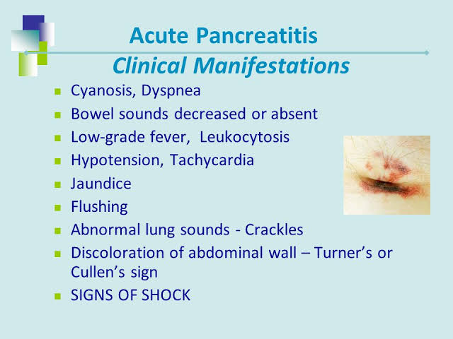


1. There is sudden and very painful mid epigastric pain and radiate to back. It hurts worst when lying in supine position. Pain becomes after drink alcohol or eating fatty meal.

2. Patient may have fever due to infection , increased heart rate and decreased BP.

3. Increase in glucose level and also amylase and lipase level.

4. Cullen's sign- there is bluish discoloration around umbilicus due to leaking of enzymes into umbilicus area.

5. Grey turner's - it is bluish discoloration of the flank area.


Treatment-


Care of patient with pancreatitis-

 Goal should be rest to pancreas , control pain , monitor for complications , administer medications as per ordered. Assessment of pancreatic enzymes and therapeutic diet.

1. Maintain NPO status of patient. Let pancreas rest and when sign symptoms get slow than provide liquid without fats.

2. Maintain IV hydration as prescribed.

3. Insertion of NG tube may prescribed by doctor which help to remove gastric contents , gas and help to Decrease vomiting.

4. Monitor glucose level and administer insulin as per ordered.

5. Monitor stool for greasy appearance which indicates steatorrhoea.

6. Monitor urine output and colour.

7. Administer pain medication as per ordered.
Morphine is contraindicated because it results spasms of sphinctor of Oddi.

8. Advise to patient to lean forward or sit up and not have to take supine position.

9. Administer pancreatic enzymes as per ordered which help to break protein and fat to prevent steatorrhoea.

10. Administer these enzymes with acidic foods and not administer with alkaline e.g milk, pudding and ice-cream.

Diet- 

1. Avoid alcohol or greasy fatty foods.
2. Low fats.
3. High proteins.
4. Limited sugar + refined carbs and complex carbs.


Friday, 19 October 2018

What is liver cirrhosis symptoms and treatment.

Liver cirrhosis-

Liver cirrhosis is a disease of the liver where liver cells becomes severely damaged and replaced with fibrous tissue and leads to scarring of the liver cells.


What causes liver cirrhosis-


1. Liver cirrhosis is most commonly caused by alcohal consumption.

2. Chronic viral hepatitis infections ( hep.A and B, E)

3. Biliary cirrhosis and bile duct blockage due to cholesterol accumulation.

4. Drugs including NSAIDS.

5. Auto immune diseases.

6. Bronze diabetes which is indicated by accumulation of Iron in the tissues included liver.

7. Wilson's disease which is genetic disorder characterized by accumulation of copper in the tissues of liver brain and other vital organs.

8. Budd chiari syndrome which is characterized by thrombus formation in hepatic veins.

9. Alpha 1 antitrypsin deficiency which is characterized by the accumulation of abnormal A1 AT in liver tissues.

10. Obesity and fat accumulation in liver tissues.

What are the normal functions of liver and what happened to it when cirrhosis occur-

1. Hepatic portal vein receive deoxygenated blood from the digestive organs. When cirrhosis occur blood can't transfer to liver and cause portal hypertension which leads to main complications called oesophageal varices.

2. Metabolization- it takes excessive amount of glucose and stored as glycogen. And also convert glycogen to glucose. When liver cirrhosis occur it leads to decrease storage of glucose and that free glucose appears in blood cause hepatic diabetes.

* Biproduct of protein breakdown is ammonia liver will take it and covert amonia into urea.
When cirrhosis occur it can't convert it into urea and free Amonia start to accumulate in brain cause severe complication called hepatic encephalopathy also amonic smell in breath.

3. Storage - liver store  Vit.B12 , A , C , E , D , k glycogen and Iron. When cirrhosis occur it leads to low vit. D and cause fractures and deficiency of vit. K leads to low coagulation.

4. Digestion - hepatocytes produces bile. That bile help in digestion and stored in gall bladder. There is bilirubin present in bile. Bilirubin in bile produces by the process when kuffer cells of liver remove old RBCs and breakdown of he hemoglobin in heme+globin. Heme is taken by hepatocytes and metabolise heme into ferrous ions and bilirubin. This bilirubin release into bile and secret through stool outside the body. Colour of stool is due to that bilirubin also called stercobilin.

When cirrhosis occur color of stool becomes clay colour stool. This bilirubin get release in blood and stored on skin and skin appear yellow in color.

5. Production of blood plasma proteins-

* Albumin- it is made by liver and maintain oncotic pressure and help to hold water inside the blood. When there is cirrhosis than no albumin lead to spread of water to interstitial space and cause Ascitis which is characterized by swelling of abdomen. There is also swelling of lower legs.

* Fibrinogen and prothrombin-
These protiens helps to clotting abilities and if cirrhosis occur , deficiency of these proteins cause low coagulation and profuse bleeding may occur to patient.
  
6. Detoxification-
Liver makes drugs less harmful to body and remove toxins. Also make alcohal less toxic.
Also remove hormone secreted by the glands. When cirrhosis occur there is decrease metabolism of estrogen which leads to get accumulate in blood. In male symptoms include gynaecomastia ( increase size of breast) and spider angiomas on chest and Arms.

Complications-

1. Portal hypertension due to narrowing of portal veins and leads to increased pressure. Increase in pressure cause 

  •  Enlarged spleen. Spleen stored WBCs and platelets which get decrease.
  •  Esophageal varices which is characterized by dilation of vessels of esophagus dur to increased pressure and get rupture.

2. Fluid volume overload due to decrease albumin and cause Ascitis and swelling in lower legs.
3. Jaundice.

4. Renal failure ( hepato renal syndrome)

5. Hepatic encephalopathy due to accumulation of ammonia in brain. Characterized by disturbed mental status.

6. Liver cancer.

7. Bone fractures.
8. Diabetes.
9. Astrexis which is characterized by flapping of hand.

Sign and symptoms-

  • Tremors 
  • Hepatic foetor.
  • Yellowish eyes and skin.
  • Loss of appetite
  • Increased bilirubin and Amonia.
  • Esophageal varices.
  • Swelling of legs and abdomen.
  • Bleeding and increase risk of infections.
  • Itchy skin
  • Confusion / coma
  • Renal failure 
  • Enlarged breast in men.
  • Flapping of hands.

Treatment-

1. Liver transplantation.
2. Shunt surgery.
3. Diuretics , beta blockers , Nitrates and other drugs prescribed according to cause of disease.
4. Blood products and vit.K for clotting.
5. Paracentesis ( removal of fluid from peritoneum layer of stomach)

Care of patient-

1. Monitor for hyper and hypoglycemia.

2. Monitor for PT/INR because patient may have risk for bleeding.

3. Check for esophageal varices and avoid coughing , vomiting , and alcohol intake.

4. Low protein diet if patient have hepatic encephalopathy and if not than provide  lean protein diet , low sodium (sodium cause water retention) ,No raw sea food , fluid restrictions ( to prevent more swelling) and oral vitamins.

5. Assessment of skin for breakdown.
6. Do not allow patient to sleep in supine position because it may lead to pressure exerted on on respiratory system and may have breathing difficulty and activity intolerance.

Preventive measures-

  • Avoid alcohol.
  • Loss weight if patient is fatty.
  • Take regular exercise
  • Practice good hygiene to reduce chances of infections.
  • Take well balanced diet. And take low salt in diet.
Take proper immunization for hepatitis infections. 


What is peptic ulcer disaese and what are preventive measures.

Peptic ulcers-


Peptic ulcers form In the upper Gastrointestinal tract that affect lining of stomach (gastric ulcers), duedenum which is part of small intestine (duedenal ulcers) and also in lower part of oesophagus.


Causes-


1. H.pylori (helicobacter pylori) it is most common cause of peptic ulcer.
2. Non steroidal anti-infalamatory drugs.(NSAIDS)
3. Mental stress.
4. Smoking.
5. Alcohol consumption.
6. Genetics.

How they form? (Pathophysiology)-


In our stomach there is mucosa layer which play important role in protection of stomach from erosion.

Mucosa-

This is layer which release mucous rich in bicarbonate and help to protect lining from acid secreted by parietal cells of stomach.
Different cells function-

1. Parietal cells release HCL acid which is helpful in digestion of food but due to its acidic nature it tend to erode mucosal line when there is low bicarbonate secretion by mucosa.

2. Chief cells release pepsinogen which mix with HCL and form pepsin.

3. G- cells release gastrin.

Prostaglandins-

These have major role in regulating perfusion in stomach which leads to good work of cells. It also regulate mucus to release bicarbonate and also control acid production from parietal cells.
This all systems of normal functions get disturbed due to all causative agents by following process- 
1. H.pylori which is most common bacteria cause erosion of lining of stomach 
When cell get damage  leads to release of histamine which give signal to parietal cells to produce more HCL and which erode stomach lining.
H.pylori is spiral shape bacteria which help it to easily enter into mucosa.

How H.pylori survive in acidic nature of stomach-

H.pylori produces urease and  breakdown to urea which produces Amonia that's why patient with peptic ulcers have amonic smell in their breath. This amoniA neutrilize acid and erode lining. This bacteria spread through contaminated water and feco-oral route.

2. NSAIDS-  these included Ibuprofen, Aspirin , Indomethacin. These drugs lead to decrease production of prostaglandins and before we studied functions of prostaglandins. These functions get disturbed and leads to erosion of lining.

Another cause also lead same process of erosion.

What sign and symptoms patient have -


Commonly sign and symptoms in this includes 
* Indigestion.
* Epigastric pain.
* Pain will be dull or aching.
* Epigastric pain.

 But there is difference between the gastric and duedenum ulcers sign and symptoms that is-

In gastric ulcers-

1. Food makes pain worst because when we take food it's enter firstly in stomach and HCL starts release which make pain worse within half an hour after ingestion of food.

2. Patient not complain pain at night.

3. Pain is dull and aching.

4. Weight loss. Because patient will be unable to take proper diet due to pain.

5. Severe vomiting with coffee , ground or bright red blood.


Duodenal ulcers-

1. Food make pain better. But pain get after 3-4 hrs after taking meal because food get into intestine after 3-4 hrs.

2. Patient complaint pain at night.

3. Pain is Inawing.

4. Weight is normal.

5. Dark colour stool due to blood present in stool.( Melena).

Diagnosis-

1. Endoscopy
2. Barium swallow.
3. CT scan.
4. Blood and stool test for H.pylori.
If H.pylori present than there will be smell of Amonia in breath.so breath test should done also carbon dioxide measurement done because urea break into Amonia and Carbon dioxide.

Complications-

1. GI bleeding.
2. Bowel blockage due to chronic obstruction.
3. Increase risk of GI cancer.

Treatment-

Medications-

* Proton pump inhibitors.
* Histamine receptor blocker.
* Antibiotics.
* Antacids.
In severe cases patient require surgery included-
1 . Vagactomy - Removal of vagus Nerve.
2. Pyloroplasty- surgery performed to widened lower part of stomach called pylorus.
3. Total gastrectomy- removal of stomach and anastomosis of oesophagus with intestine. After this it's important to look for dumping syndrome which is characterized by the emptying of  food directly into intestine in bulk and cause many complications like sudden increase in sugar etc.

Education to patient-

1. Eat small and frequent food.
2. Do not drink fluid with food.
3. Avoid sugary food and drinks.
4. Avoid very hot and cold food.
5. Take high protein , high fibers and low carb food.
6. Avoid spicy food , coffee, alcohol and fried foods.
7. Encourage to consume  food easily digestible eg. Rice and banana.


Thursday, 18 October 2018

Importance of Urinary Catheter Care


Urinary catheter-It is a tube like structure made up of latex, polyurethane and silicon.This is inserted into bladder of patient via urethra.It allow drainage of urine from bladder freely when urine retention occurred and patient is unable to pass urine.Another purposes also to make diagnosis of any urinary related disease.It may prescribed by doctor for short and long duration according to the patient condition.

 Why it is important to care of patient with catheter?-

Most of patients in hospital acquire another bacterial infections along with present illness.
Main factors for this includes sometimes wrong hospital practices and procedures and also care provided to the patient in home.patient and attendent should know regarding all procedures and treatment regimens.Urinary catheter is most important cause of another bacterial infection.That attain more awareness to provide perfect care after and during insertion.
In hospital setting according to protocols it's responsibility of nursing officer to provide care in her  three shifts of day. 

1.In morning shift around 9-10Am.
2.In evening shift around 2-3pm.
3.In night it's in two times.first around 9-10 pm and second around 5-6am

When patient is in home care settings same timings should follow.
Infections caused by urinary catheter is called catheter associated urinary tract infection(CAUTI Bundle)

Steps of catheter care in female patient-

1. Gather all supplies and equipments.

 *  Two big towels
 *  One small clean towel or washing cloth.
 *  Basin of water. Temperature of water       should maintained according to patient   comfort.
 *  Soap
 *  Gloves

2. Provide privacy to patient and adjust bed as patient and your own comfort.
3. Wash hand with soap and dry well.
4. Spread one big towel under perineal area to avoid bedsheets contamination.
5. Wear gloves.
6. Wet cloth or small towel with water and apply soap on it.
7. Seprate thighs well to expose perineal area well and clean perineal area gently. Open meatus with one hand and hold catheter near the meatus to prevent pulling off of catheter. with the help of another hand apply washcloth on catheter closets to meatus and wipe gently tube downward in one direction. Change site of wet cloth with each wash and repeat 4-5 times or more.
8. Take another dry towel and wipe wet area with it.
9. Discard water in basin and gloves carefully.

In male patients- 

All equipment are same used in female patient and procedure also same unless-
1. Hold penis straight upward with one hand and with another hand apply wet cloth with soap and clean gently area and skin around the catheter. Also clean penis and scrotum well.
2. Clean catheter for 4inch from insertion site.

These practices can prevent patient from any complications and another infections to occur. It is very important to provide care in regular manner if you skip than infection may occur.