Tuesday, 30 October 2018

Asthma and it's treatment

Asthma is chronic lung disease that cause narrowing and inflammation of the airway.
It effect bronchi and bronchioles and these are chronically inflamed.

There are smooth muscles at outer part of bronchioles which constrict and dilated . When Asthma attack occur this leads to constriction of muscle and cause chest tightness and dyspnea.

Inside the bronchioles there are goblet cells  which are help to produce mucous. When Asthma occur they strat over stimulate  and start to produce extra mucous wnich lead to decreased air flow.

Causes-


1. Smoke , pollen , pollution , perfumes.

2. Danders , dust mites , pests.

3. Cold and dry air .

4. Respiratory infection.

5. Exercise induced.

6. Drugs e.g. badrenergics  blockers e.g. NSAIDS  , Aspirin.


Early sign and symptoms-


1. Shortness of breath.

2. Easily fatigued with physical activity.

3. Frequent cough at night.

4. Symptoms related to cold , sneezing , scratchy throat.

5. Irritable.

Active sign and symptoms-


1. Chest tightness.
2. Wheezing.
3. Coughing.
4. Dyspnea.
5. Increased pulse rate.

And if these signs are not treated than it can lead to -

1. Inhaler can't work.
2. Patient can't speak.
3. Chest refraction.
4. Cyanosis mainly on lips skin.
5. Sweaty and need medical treatment very fast.

Treatment-


Medications-


1. Bronchodilators- 


A. Short acting- Albuterol. Fast relief during an attack. And not used for daily treatment. These medications should not use more than 2 time for week.

B. Long acting - a. Salmetrol 

                             b. Symbicort

These both drugs used with corticosteroids. And not used for acute attack.

C. Anticholinergics-  inhaled and are short acting ( Ipratropium).

Long actingTiotropium . This medication cause dry mouth.

D. Theophylline - oral and not common used because cause toxicity.

2. Anti- inflammatory- 


a. Corticosteroid - Inhaled , Intravenous , oral , and used for long term   not used in acute attack. Fluticasone , Budesonide , Beclomethosone.  They cause thrush and to prevent this use spacer with it.

- These are used after 5 mintues of bronchodilators if bronchodilator are prescribed along with this.

- These corticosteroid may lead to osteoporosis and catract.

b. Leukotriene modifiers- these are oral medications .

 Montelukast- these blocks the functions of of leukotriene (smooth muscles to constrict and Increased mucous production). These relaxed smooth muscles and decreased mucous. These are not used in acute attack.


c. Omalizumab - These blocks role of immunoglobulin IGE and decrease allergic response. These drugs are used when Asthma is poorly controlled and when another treatment are not working. Its not for quick relief and no live vaccine should used with it.

d. Cromolyn ( Inhaled) - non steroidal anti- allergy . Decreased functions of cells secreting histamine and also used for long term not for quick relief.
 Patient may have sneezing , burning in nose , watery eyes , bad taste in mouth.

Self care- quit smoking and avoid substance that cause allergy.

Supportive care - oxygen therapy ( provide extra oxygen to patient have difficulty in breathing  as per ordered ).

Sunday, 28 October 2018

What is diverticulitis


Diverticulitis is formation of hollow sac cavities through intestinal wall. Found throughput the large intestine and most common in sigmoid.

Person can have many out pouching area is diverticula and or single called diverticulum.
When diverticulitis occur there is formation of hollow sac from diverticulum.

Patient with disease is usually asymptomatic and sometimes get experience disturbed bowel pattern and bloating.

Causes-


Causes are not 100% known .

- low fiber diet.
- constipation and cause Increased pressure on wall cause herniation.

Diagnosis-

1. Colonoscopy-  tube inserted inside the colon through rectum with camera and visualised on screen.

2. Barium anema- It is X- Ray examination in which a contrast with barium metal element delivered into the colon. And X- Ray taken of area where this barium moved and give pictures if obstructions occur.

3. CT scan of abdomen.

Complications- 

1. Painless bleeding , blood with stool. Rupture of diverticula leads to leak of the content in abdomen which may cause peritonitis.

2. Strictures and obstructions.

3. Formation of fistula ( abnormal connection between organ and other organ ). Most common fistula occur from intestine to the bladder are most common which leads to the leakage of all content and bacteria into the bladder.

4. Mainly this disease is supposed to cause by entering of stool ito the pouch of intestine which may cause inflammation and other cause will be the eating of seed food. So these food have to avoid.

5. Other cause are Increased pressure to the colon that lead to the ruptures of diverticula and invade by bacteria cause abscess formation.

6. Abscess formation may lead to peritonitis and obstruction and lead to pain , fever and Increased wbc count.

Sign and symptoms-


1. Pain in left lower quadrant.

2. Bloating of stomach.

3. Unrealting cramps - intense pain like the child birth pain of 6 child.

4. Constipation or diarrhea.

5. High temperature due to infection.

Treatment-


1. Drain abscess of infected pouch.

2. In mild cases oral antibiotics and bowel rest ordered by doctor.

3. In moderate case I.V antibiotics , hospital stay , IV fluid and bowel rest ordered.

4. In cronic cases bowel resection may need multiple surgeries where can't may be recommend.

Patient care-


1. Focus on GI assessment.

2. Administer IV antibiotics in the severe cases and oral if mild cases. 

3. Diet should maintain NPO ( mill per oral ) and administer IV fliud as per ordered.

4. Nurse have to monitor sign and symptoms of peritonitis for unrelating fever , Increased pain , incIncrea pulse rate , Increased Heart rate , Increased bloating and hydration.

5. When symptoms recover as per the the doctor order strat clear liquid , broaths , pulp free clear juice.

6. Low fiber diet should provide to patient to work intestine in good work. Diet included white rice , cooked / skimmed fruits , vegetables , egg and no high fibers.

7. When patient get improved than have to provide high fiber diet to avoid constipation. Diet included fresh veg , fruits , grains , beans , plenty of fluid 2-3 l per day.

8. As per doctor ordered administer psyllium which absorb water from intestine and help to pass stool easily.

9. Probiotic also help to maintain health flora.


Saturday, 27 October 2018

Appendicitis and it's treatment


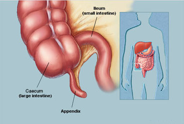
Appendicitis is the inflammation of the  appendix. Appendix is the finger like structure originated from the cecum part of ascending colon.

Appendix functions-

Appendicitis is the inflammation of the appendix . Appendix play an important role in storing good bacteria in the GI tract after a diarrhea and illness and help to maintain gut flora.

Causes-


1. Obstructions- Fecolith ( term used for calcified and obstruction and lead to rupture ) which cause perforation , Abscess formation peritonitis.

2. Parasites , foreign bodies , swollen lymph nodes in lining of appendix.

3. Trauma and injuries.

Pathophysiology-

1. Blockage in the lumen of appendix leads to increase pressure inside the appendix because of increase amount of mucous from mucosal lining and increase fluid along with Increased bacteria. These bacteria are already present in appendix which maintain flora. These bacteria can go anywhere and increase again pressure so within 48-72 hrs patient have risk of perforation.

2. When Increased pressure cause major venous obstruction ( occlusion of the blood flow) and blood remain stagnant which leads to clot formation and cause ischemia to appendix ( diminished blood supply to appendix) which cause slow breakdown of walls of appendix.

3. These all conditions lead to the peritonitis and which leads to septic shock and death.

Sign and symptoms-


1. Abdominal pain which is dull around the belly button and radiate right Lower quadrant also called mecburney's point.

2. Poor appetite.

3. Elevated temperature.

4. Nausea and vomiting.

5. Desire to be in lie in fetal position which give relief in pain.

6. Increased wBCs and infection.

7. Inability to pass gas , stool and may have constipation and diarrhea.

8. Experience rebound tenderness i.e. when press there will be no pain but when left pressure there is too much pain.

9. Abdominal rigidity.

Treatment-


Surgeries- 

1. Appendectomy - Removal of Appendix.

2. Laparotomy - surgical opening made in abdomen to treat and diagnose many diseases.

3. Laproscopic surgeries- A vedio camera with tube is inserted in small cut on body to repair and remove tissues.

Medications-

Generally antibiotics are given to patient to overcome the infection and reduce infection.

Patient care-


Pre- op ( before operation)-

1. Vital signs monitor.

2. Monitor sign of peritonitis and perforation.

Perforation-
 
In this case patient have relief of pain with Increased level of pain. If pain get relief after the sever pain it will indicate that appendix may have rupture and all content get release into the abdomen. Which leads to the peritonitis and cause.

  • Increase heart rate.
  • Increase pulse rate.
  • Increase temperature.
  • Pain relief with fetal position.
3. Do not provide hot applications on stomach to pain relief because it can lead to rupture of appendix.

4. Do not use enema and laxatives because it can lead to rupture of appendix.

Post op care ( after operation )-

1. Monitor vital sign.

2. Monitor site for infection.

3. Care for drain as per doctor ordered.

4. Positioning should provide to the patient to remove drainage.

5. Ambulate patient which help to come out all waste fluid easily into drain.

6. Provide spirometry , coughing and deep breathing exercises.

7. Administer IV antibiotics as per ordered.

8. Administer pain medications as per ordered.

9. Monitor bowel sound and should present 2- 3 days after surgery.

10. Also flatus should pass within 2 days.


Thursday, 25 October 2018

Crohan's disease and it's treatment.


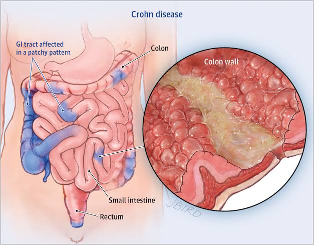
It is a type of inflammaory bowel disease that cause inflammation ulcers formation in the GI tract . Another type of inflammaory bowel disease is ulcerative colitis.

Some common facts about the disease-


1. This disease can be found in both large and small intestine but ulcerative colitis occur in only large intestine.

2. It most likely found in the terminal of ileum and starts from the colon.

3. This disease affect all lining of the bowel and ulcerative colitis only effect the inner lining of the bowel it cause more complications.

4. Diseases found in scattered patches of infected lines after healthy lines this called cobble stone appearance.

5. Causes of diseases are unknown and most commonly caused by the faulty immune system and may be triggers by the genes.

6. This diseases have periods of flare up and remission.

7. There is no care and no colostomy and ileostomies because all parts of small and large intestine are affected. Only bowel resection , medication and diet can prevent this.

Types -


Ileocolitits - inflammation found in ileum colon.

Gastroduodenal- ulcers found in part of colon of stomach and duodenum.

Jejuno ileitis - found in jejunum.

Ileitis- ulcers found in the ileum.

Granulomatous colitis - this is only found in the colon.

Causes- 

Causes are unknown but it is mainly caused by immunodeficiency ,  genetic and environmental factors.

Complications- 

1. Abscessing fistulas - it forms sepsis. Abscessing packets of infection form within the intestine wall. Fistulas ulcers and abscess form deep in intestine wall creating an opening channel and passage of infection between intestine to intestine , intestine to organ and other skin surface.

Entro enteric- intestine to intestine abscess formations.

Perianal - due to anal abscess.

Entro vesical - abscess form Intestine to bladder.

Entrovaginal - abscess form intestine to vagina 

Entro-cutaneous -  abscess form intestine to skin surfaces.

2. Malnaurisment-

Small intestine get infalmed and all enzymes required for digestion of food not secret and cause malnaurisment.

3. Anal tear and fissures is complication.

4. strictures- 

obstructions is common complications major narrowing of intestinal wall due to chronic inflammation which leads to scarring and limit flow of GI contents , food get stuck and cause obstructions.

5. Other complications will the arthritis , stress in gall bladder , skin rashes and eyes issues.

Sign and symptoms- 


1. Abdominal pain and mainly in the right Lower side because it mainly effect ileum and that part is present in the right Lower side.

2. Ulcers in mouth and GI tract.

3. Diarrhea may be with pus , blood and mucous.

4. Loss of weight , malnaurisment  and electrolyte imbalance.

5. Fissure Anal that bleed .

6. Bloating.

Treatment-


Medications-


NSAIDS - decrease pain decrease inflammation and fever.

  Ant inflammatory - prevent swelling in tissues

Immuno suppressors- reduce immune response.

Steroids - modifies and stimulate hormonal effect.

Analgesic - relief pain.

Surgery-

Colostomy - surgical creation of abdominal opening In large intestine where stool can leave.

Ileostomy- surgical connecting of small intestine to opening in abdomen so stool can leave to it.

Proto colectomy- surgical removal of rectum and all part of colon.

Colectomy- surgical removal of all part of colon.

Iv fliud therapy-

Administration of Iv fluids done to recover electrolyte imbalance.

Enema may be administer to increase bowel movement and clear out stool .

Patient Care-

Goal -

Help patient to understand diet medications , surgeries .

1. Advice for smoke cessation.

2. In severe cases have to give rest for bowel and maintain NPO and administer TPN as per ordered. Patient may have risk for nutrition imbalance.

3. Daily weight monitoring.

4. Monitor intake and output.

5. GI assessment eg. Frequency of bowel movement have to check and also bowel sound.

6. Patient have to advise to avoid food with high fiber  , dairy products, spicy food fatty food .

7. Consume high protiens.


Tuesday, 23 October 2018

Ulcerative colitis and its treatment


A type of inflammatory bowel disease that causes inflammation ulcers in the inner lining of the colon rectum.

Causes-

1. Autoimmune triggering the body to inflame the colon.
2. High fat diet.
3. Milk allergy
4. Stress and illness.
5. Viral and bacterial infection.
6. NSAIDS

Types of ulcerative colitis-


1. Ulcerative proctitis- affect the area of rectum.
2. Proctus sigmoiditis- affect the area of rectum and sigmoid.
3. Left sided colitis- it affect rectum sigmoid and descending.
4. Pancolitis- sever case affect all colon.

What happened to large intestine when ulcer forms- (pathophysiology)-

Inflammation in large intestine leads to the cell damage and kill cells of the lining. Which leads to the ulcer formation. These ulcer formation cause the bleeding , pus formation which leads to the fever , diarrhea and anemia due to blood loss. Patient have abdominal pain due to inflammation. Colon can't do it's function of absorption of water which may lead to the dehydration, fluid electrolytes imbalance and decreased appetite and leads to weight loss.
Patient have frequent bowel movements so feel urgent frequency.

Sign and symptoms-


1. Urgent bowel movements.

2. Low RBCs cause anemia .

3. Weight loss due to improper digestion of food.

4. Cramps in abdomen which are very painful. This is due to inflammation and after taking food this pain becomes more severe.

5. Electrolyte imbalance occur due to dehydration because large intestine fail to absorb water.

6. Fever due to pus formation and infections.

7. Rectal bleeding.

8. Severe diarrhea with blood pus mucosa.


Complications-


1. Scars in the lining get flare up again after healing that cause  pseudo polyps, scar tissue and narrowing.

2. With sever case large intestine starts to loose it's pouch like structure called hustra which help to chum food properly and leads to colon become smooth called lead pipe sign.

3. Rupture of intestine due to hole formation after ulcers leads to leakage of bacteria , pus into abdominal cavity which leads to peritonitis.

4. Toxic megacolon occur due to the overwhelming inflammation which dilate colon and that become paralyzed and can't work properly. Which is very life threatening condition.

5. Patient is also in risk of colon cancer.

Diagnosis- 


1. Colonoscopy
2. Barium enema.

Treatment- 

1. Surgery in severe cases including.

Proctocolectomy - surgical removal of rectum and all part of colon.

Ileostomy - surgical opening of ileum.

Ileoanal Anastomosis with J pouch - in this rectum removal and pouch create that attached to ilium which allows stool to pass from small intestine to Anus without Ostomy.

2. Medications- 


A . Anti inflammatory - 5 Amino salicylate (sulfate salazine) most important and these are first line treatment and can't given to patient have allergy with sulfa.

B. Corticosteroid (predinsone) these are not used long term and have trapped off because have side effect e.g Increased glucose level , thinning of skin , bruising and osteoporosis.

C. Immuno suppressors- these are strong drugs and are used in severe cases of ulcerative colitis. Common drug is Azathiarine. ( Imuron)
These drugs decreased immune system and have great risk to development of the cancer.

D. Immuno modulators ( Adalimumab, Humaria).- 
They blocks TNF which is protien play role in inflammation.
 

Diet- 


Patient care-

1. Monitor vital signs .

2. Monitor bowel movement , sounds and 
frequency.

3. Focus to complications e.g . Peritonitis , toxic megacolon , abdominal pain , bloating , fever, increased heart rate , Increased respiratory rate .

4. Keep NPO and administrator IV fluid.

5. Food not given included high fiber because they require lot of digestion , nuts , popcorn , raw fruit and vegetables. Dairy , spicy and high fat food.

6. Easy to digest food includes low fiber high protien diet.

7. Regular colon cancer screening should done.

8. Ostomy care should provide to patient.


Saturday, 20 October 2018

Pancreatitis and treatment

Pancreatitis-


Pancreatitis is the inflammation of pancreas. In pancreas there are number of cells that produces enzymes , hormones and when inflammation occur it leads to digestion of the pancreas by it's own enzymes or cause irreversible structural damage to the organ.


Pancreatitis is of two types-

1. Acute pancreatitis
2. Chronic pancreatitis.

What are the functions of pancreas and how they get affected with pancreatitis-

Pancreas have two types of functions-

1. Exocrine by Acinar cells-

It secret inactive enzymes into pancreatic duct these enzymes are-

  • Amylase - breakdown carbohydrates to glucose
  • Protease -  breakdown protiens.
  • Lipase    - breakdown fats
Pancreas have epithelial cells which release bicarbonate.

2.  Endocrine Islets of Langerhans-

It release insulin , glucagon , somatostatin , pancretic polypeptide into blood stream.

These all enzymes are not activated and they become activated when enter into the duodenum which is part of small intestine. These enzymes reach to duodenum via pancreatic duct.


When pancreatitis occur  which cause inflammation and pancreas get swell up. Enzymes become unable to release due to obstruction in path and these enzymes starts to leak surrounding the pancreatic tissues and also in surrounding organs which leads many complications.

Complications included-


1. Chronic pancreatitis leads to damage to isletI of Langerhans and cause low production of insulin which cause diabetes mellitus.

2. Malabsorption   occur due inadequate secretion of enzymes and their release into small intestine. Which leads to improper digestion of food and cause malabsorption.

3. GI pain due to inflammation of tissues.

4. Internal bleeding due to the inflamation 
of blood vessels.

5. Ascitis occur due to leaking of pancreatic secretions into peritoneum.

6. Fibrosis

7. Cyst formation.

8. Respiratory distress.

What Is difference between acute and chronic pancreatitis? -

Acute pancreatitis-


1. Sudden inflammation due to something that triggers the digestive enzymes to activate inside the pancreas.

2. These activated enzymes inside the pancreas starts to digest cells of pancreas. Which leads to swell up of pancreas and inflamed tissues died cause fibrosis , cyst formation  and abscess formation.

3. Due to location of pancreas it leads to inflammation of other organs. E.g respiratory distress etc.

Chronic pancreatitis-


1. Chronic pancreatitis is repeated episode of acute pancreatitis but most common cause is years of alcohol abuse.

2. It is irreversible change to pancreas.

3. There is complete Loss of endocrine and exocrine cells functions.

4. Complete damage to pancreatic ducts functions.

What are causes of acute and chronic pancreatitis? -


Acute pancreatitis- 

Mainly two causes are dominant-

1. Gallstones- hardened deposit of undissolved cholesterol salt or bilirubin that can block common bile duct cause reverse transport of enzymes. These enzymes get activated and start to digest it's own cells in pancreas.

2. Alcohal - leads to damage of the cells of pancreas (Acinar cells) , pancretic ducts and lead to the activation of enzymes.
Other-

  • Abdominal trauma.
  • Medications.
  • Infections.
  • Tumors.
  • Genetics.
  • High triglycerides level.
  • High calcium level.

Chronic pancreatitis -


1. Long term alcohal abuse- recurrent inflammation and damage to the structure of pancreas occur which leads to the chronic pancreatitis.

2. Cystic fibrosis- there is lack of CFTR proteins. This protein play important role in the movement of chloride ions and maintain balance of water and salt in epithelial cells that lining the pancretic ducts. When cystic fibrosis occur it leads to low production of bicarbonate and leads to thick mucous in pancreatic duct. This thick mucous starts blockage and leads to activate digestive enzymes inside pancreas leads to fibrosis of pancreatic tissues.

 Other - 

1. Increased calcium .
2. Increased  cholesterol.

Diagnosis-


1. Blood test for serum amylase and lipase. Also to find out increase electrolytes.
2. CT scan and Ultrasound.
3. ERCP - use to diagnose and treatment purposes. It assess pancreas , bile ducts and gall bladder.

What are sign and symptoms of acute pancreatitis?-


1. There is sudden and very painful mid epigastric pain and radiate to back. It hurts worst when lying in supine position. Pain becomes after drink alcohol or eating fatty meal.

2. Patient may have fever due to infection , increased heart rate and decreased BP.

3. Increase in glucose level and also amylase and lipase level.

4. Cullen's sign- there is bluish discoloration around umbilicus due to leaking of enzymes into umbilicus area.

5. Grey turner's - it is bluish discoloration of the flank area.


Treatment-


Care of patient with pancreatitis-

 Goal should be rest to pancreas , control pain , monitor for complications , administer medications as per ordered. Assessment of pancreatic enzymes and therapeutic diet.

1. Maintain NPO status of patient. Let pancreas rest and when sign symptoms get slow than provide liquid without fats.

2. Maintain IV hydration as prescribed.

3. Insertion of NG tube may prescribed by doctor which help to remove gastric contents , gas and help to Decrease vomiting.

4. Monitor glucose level and administer insulin as per ordered.

5. Monitor stool for greasy appearance which indicates steatorrhoea.

6. Monitor urine output and colour.

7. Administer pain medication as per ordered.
Morphine is contraindicated because it results spasms of sphinctor of Oddi.

8. Advise to patient to lean forward or sit up and not have to take supine position.

9. Administer pancreatic enzymes as per ordered which help to break protein and fat to prevent steatorrhoea.

10. Administer these enzymes with acidic foods and not administer with alkaline e.g milk, pudding and ice-cream.

Diet- 

1. Avoid alcohol or greasy fatty foods.
2. Low fats.
3. High proteins.
4. Limited sugar + refined carbs and complex carbs.


Friday, 19 October 2018

What is liver cirrhosis symptoms and treatment.

Liver cirrhosis-

Liver cirrhosis is a disease of the liver where liver cells becomes severely damaged and replaced with fibrous tissue and leads to scarring of the liver cells.


What causes liver cirrhosis-


1. Liver cirrhosis is most commonly caused by alcohal consumption.

2. Chronic viral hepatitis infections ( hep.A and B, E)

3. Biliary cirrhosis and bile duct blockage due to cholesterol accumulation.

4. Drugs including NSAIDS.

5. Auto immune diseases.

6. Bronze diabetes which is indicated by accumulation of Iron in the tissues included liver.

7. Wilson's disease which is genetic disorder characterized by accumulation of copper in the tissues of liver brain and other vital organs.

8. Budd chiari syndrome which is characterized by thrombus formation in hepatic veins.

9. Alpha 1 antitrypsin deficiency which is characterized by the accumulation of abnormal A1 AT in liver tissues.

10. Obesity and fat accumulation in liver tissues.

What are the normal functions of liver and what happened to it when cirrhosis occur-

1. Hepatic portal vein receive deoxygenated blood from the digestive organs. When cirrhosis occur blood can't transfer to liver and cause portal hypertension which leads to main complications called oesophageal varices.

2. Metabolization- it takes excessive amount of glucose and stored as glycogen. And also convert glycogen to glucose. When liver cirrhosis occur it leads to decrease storage of glucose and that free glucose appears in blood cause hepatic diabetes.

* Biproduct of protein breakdown is ammonia liver will take it and covert amonia into urea.
When cirrhosis occur it can't convert it into urea and free Amonia start to accumulate in brain cause severe complication called hepatic encephalopathy also amonic smell in breath.

3. Storage - liver store  Vit.B12 , A , C , E , D , k glycogen and Iron. When cirrhosis occur it leads to low vit. D and cause fractures and deficiency of vit. K leads to low coagulation.

4. Digestion - hepatocytes produces bile. That bile help in digestion and stored in gall bladder. There is bilirubin present in bile. Bilirubin in bile produces by the process when kuffer cells of liver remove old RBCs and breakdown of he hemoglobin in heme+globin. Heme is taken by hepatocytes and metabolise heme into ferrous ions and bilirubin. This bilirubin release into bile and secret through stool outside the body. Colour of stool is due to that bilirubin also called stercobilin.

When cirrhosis occur color of stool becomes clay colour stool. This bilirubin get release in blood and stored on skin and skin appear yellow in color.

5. Production of blood plasma proteins-

* Albumin- it is made by liver and maintain oncotic pressure and help to hold water inside the blood. When there is cirrhosis than no albumin lead to spread of water to interstitial space and cause Ascitis which is characterized by swelling of abdomen. There is also swelling of lower legs.

* Fibrinogen and prothrombin-
These protiens helps to clotting abilities and if cirrhosis occur , deficiency of these proteins cause low coagulation and profuse bleeding may occur to patient.
  
6. Detoxification-
Liver makes drugs less harmful to body and remove toxins. Also make alcohal less toxic.
Also remove hormone secreted by the glands. When cirrhosis occur there is decrease metabolism of estrogen which leads to get accumulate in blood. In male symptoms include gynaecomastia ( increase size of breast) and spider angiomas on chest and Arms.

Complications-

1. Portal hypertension due to narrowing of portal veins and leads to increased pressure. Increase in pressure cause 

  •  Enlarged spleen. Spleen stored WBCs and platelets which get decrease.
  •  Esophageal varices which is characterized by dilation of vessels of esophagus dur to increased pressure and get rupture.

2. Fluid volume overload due to decrease albumin and cause Ascitis and swelling in lower legs.
3. Jaundice.

4. Renal failure ( hepato renal syndrome)

5. Hepatic encephalopathy due to accumulation of ammonia in brain. Characterized by disturbed mental status.

6. Liver cancer.

7. Bone fractures.
8. Diabetes.
9. Astrexis which is characterized by flapping of hand.

Sign and symptoms-

  • Tremors 
  • Hepatic foetor.
  • Yellowish eyes and skin.
  • Loss of appetite
  • Increased bilirubin and Amonia.
  • Esophageal varices.
  • Swelling of legs and abdomen.
  • Bleeding and increase risk of infections.
  • Itchy skin
  • Confusion / coma
  • Renal failure 
  • Enlarged breast in men.
  • Flapping of hands.

Treatment-

1. Liver transplantation.
2. Shunt surgery.
3. Diuretics , beta blockers , Nitrates and other drugs prescribed according to cause of disease.
4. Blood products and vit.K for clotting.
5. Paracentesis ( removal of fluid from peritoneum layer of stomach)

Care of patient-

1. Monitor for hyper and hypoglycemia.

2. Monitor for PT/INR because patient may have risk for bleeding.

3. Check for esophageal varices and avoid coughing , vomiting , and alcohol intake.

4. Low protein diet if patient have hepatic encephalopathy and if not than provide  lean protein diet , low sodium (sodium cause water retention) ,No raw sea food , fluid restrictions ( to prevent more swelling) and oral vitamins.

5. Assessment of skin for breakdown.
6. Do not allow patient to sleep in supine position because it may lead to pressure exerted on on respiratory system and may have breathing difficulty and activity intolerance.

Preventive measures-

  • Avoid alcohol.
  • Loss weight if patient is fatty.
  • Take regular exercise
  • Practice good hygiene to reduce chances of infections.
  • Take well balanced diet. And take low salt in diet.
Take proper immunization for hepatitis infections. 


What is peptic ulcer disaese and what are preventive measures.

Peptic ulcers-


Peptic ulcers form In the upper Gastrointestinal tract that affect lining of stomach (gastric ulcers), duedenum which is part of small intestine (duedenal ulcers) and also in lower part of oesophagus.


Causes-


1. H.pylori (helicobacter pylori) it is most common cause of peptic ulcer.
2. Non steroidal anti-infalamatory drugs.(NSAIDS)
3. Mental stress.
4. Smoking.
5. Alcohol consumption.
6. Genetics.

How they form? (Pathophysiology)-


In our stomach there is mucosa layer which play important role in protection of stomach from erosion.

Mucosa-

This is layer which release mucous rich in bicarbonate and help to protect lining from acid secreted by parietal cells of stomach.
Different cells function-

1. Parietal cells release HCL acid which is helpful in digestion of food but due to its acidic nature it tend to erode mucosal line when there is low bicarbonate secretion by mucosa.

2. Chief cells release pepsinogen which mix with HCL and form pepsin.

3. G- cells release gastrin.

Prostaglandins-

These have major role in regulating perfusion in stomach which leads to good work of cells. It also regulate mucus to release bicarbonate and also control acid production from parietal cells.
This all systems of normal functions get disturbed due to all causative agents by following process- 
1. H.pylori which is most common bacteria cause erosion of lining of stomach 
When cell get damage  leads to release of histamine which give signal to parietal cells to produce more HCL and which erode stomach lining.
H.pylori is spiral shape bacteria which help it to easily enter into mucosa.

How H.pylori survive in acidic nature of stomach-

H.pylori produces urease and  breakdown to urea which produces Amonia that's why patient with peptic ulcers have amonic smell in their breath. This amoniA neutrilize acid and erode lining. This bacteria spread through contaminated water and feco-oral route.

2. NSAIDS-  these included Ibuprofen, Aspirin , Indomethacin. These drugs lead to decrease production of prostaglandins and before we studied functions of prostaglandins. These functions get disturbed and leads to erosion of lining.

Another cause also lead same process of erosion.

What sign and symptoms patient have -


Commonly sign and symptoms in this includes 
* Indigestion.
* Epigastric pain.
* Pain will be dull or aching.
* Epigastric pain.

 But there is difference between the gastric and duedenum ulcers sign and symptoms that is-

In gastric ulcers-

1. Food makes pain worst because when we take food it's enter firstly in stomach and HCL starts release which make pain worse within half an hour after ingestion of food.

2. Patient not complain pain at night.

3. Pain is dull and aching.

4. Weight loss. Because patient will be unable to take proper diet due to pain.

5. Severe vomiting with coffee , ground or bright red blood.


Duodenal ulcers-

1. Food make pain better. But pain get after 3-4 hrs after taking meal because food get into intestine after 3-4 hrs.

2. Patient complaint pain at night.

3. Pain is Inawing.

4. Weight is normal.

5. Dark colour stool due to blood present in stool.( Melena).

Diagnosis-

1. Endoscopy
2. Barium swallow.
3. CT scan.
4. Blood and stool test for H.pylori.
If H.pylori present than there will be smell of Amonia in breath.so breath test should done also carbon dioxide measurement done because urea break into Amonia and Carbon dioxide.

Complications-

1. GI bleeding.
2. Bowel blockage due to chronic obstruction.
3. Increase risk of GI cancer.

Treatment-

Medications-

* Proton pump inhibitors.
* Histamine receptor blocker.
* Antibiotics.
* Antacids.
In severe cases patient require surgery included-
1 . Vagactomy - Removal of vagus Nerve.
2. Pyloroplasty- surgery performed to widened lower part of stomach called pylorus.
3. Total gastrectomy- removal of stomach and anastomosis of oesophagus with intestine. After this it's important to look for dumping syndrome which is characterized by the emptying of  food directly into intestine in bulk and cause many complications like sudden increase in sugar etc.

Education to patient-

1. Eat small and frequent food.
2. Do not drink fluid with food.
3. Avoid sugary food and drinks.
4. Avoid very hot and cold food.
5. Take high protein , high fibers and low carb food.
6. Avoid spicy food , coffee, alcohol and fried foods.
7. Encourage to consume  food easily digestible eg. Rice and banana.


Thursday, 18 October 2018

Importance of Urinary Catheter Care


Urinary catheter-It is a tube like structure made up of latex, polyurethane and silicon.This is inserted into bladder of patient via urethra.It allow drainage of urine from bladder freely when urine retention occurred and patient is unable to pass urine.Another purposes also to make diagnosis of any urinary related disease.It may prescribed by doctor for short and long duration according to the patient condition.

 Why it is important to care of patient with catheter?-

Most of patients in hospital acquire another bacterial infections along with present illness.
Main factors for this includes sometimes wrong hospital practices and procedures and also care provided to the patient in home.patient and attendent should know regarding all procedures and treatment regimens.Urinary catheter is most important cause of another bacterial infection.That attain more awareness to provide perfect care after and during insertion.
In hospital setting according to protocols it's responsibility of nursing officer to provide care in her  three shifts of day. 

1.In morning shift around 9-10Am.
2.In evening shift around 2-3pm.
3.In night it's in two times.first around 9-10 pm and second around 5-6am

When patient is in home care settings same timings should follow.
Infections caused by urinary catheter is called catheter associated urinary tract infection(CAUTI Bundle)

Steps of catheter care in female patient-

1. Gather all supplies and equipments.

 *  Two big towels
 *  One small clean towel or washing cloth.
 *  Basin of water. Temperature of water       should maintained according to patient   comfort.
 *  Soap
 *  Gloves

2. Provide privacy to patient and adjust bed as patient and your own comfort.
3. Wash hand with soap and dry well.
4. Spread one big towel under perineal area to avoid bedsheets contamination.
5. Wear gloves.
6. Wet cloth or small towel with water and apply soap on it.
7. Seprate thighs well to expose perineal area well and clean perineal area gently. Open meatus with one hand and hold catheter near the meatus to prevent pulling off of catheter. with the help of another hand apply washcloth on catheter closets to meatus and wipe gently tube downward in one direction. Change site of wet cloth with each wash and repeat 4-5 times or more.
8. Take another dry towel and wipe wet area with it.
9. Discard water in basin and gloves carefully.

In male patients- 

All equipment are same used in female patient and procedure also same unless-
1. Hold penis straight upward with one hand and with another hand apply wet cloth with soap and clean gently area and skin around the catheter. Also clean penis and scrotum well.
2. Clean catheter for 4inch from insertion site.

These practices can prevent patient from any complications and another infections to occur. It is very important to provide care in regular manner if you skip than infection may occur.